Introduction
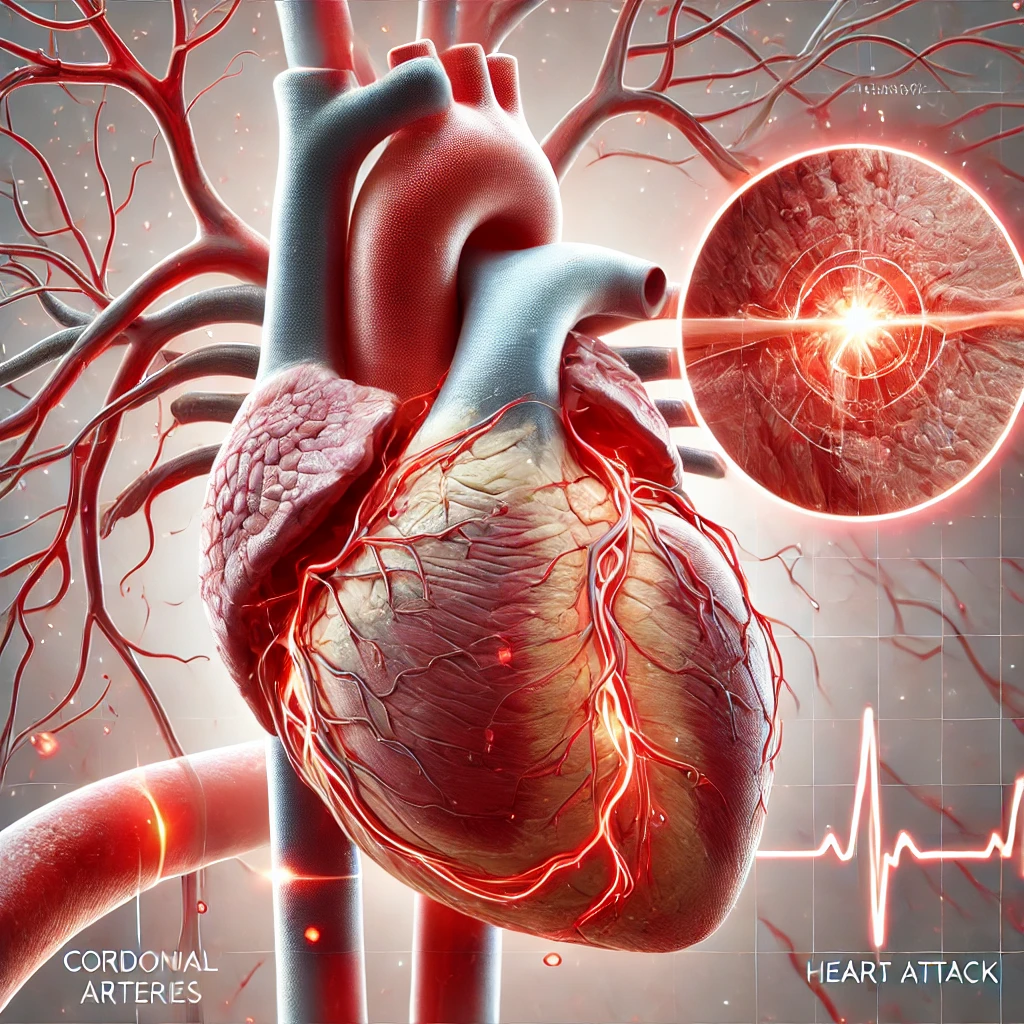
A congenital heart defect (CHD), a congenital heart defects , congenital cardiovascular malformation, or congenital heart illness, denotes abnormalities in the anatomical composition of the heart or major blood vessels that are evident from birth. A congenital heart defect is categorized as a type of cardiovascular disease. The manifestation of signs and symptoms is contingent upon the precise nature of the problem. The range of symptoms can span from being asymptomatic to posing a risk to one’s life. When symptoms are present, they exhibit variability and encompass fast respiration, bluish discolouration of the skin (known as cyanosis), inadequate weight growth, and a sense of fatigue. Coronary heart disease (CHD) is not typically associated with the manifestation of chest pain. Comorbidities do not accompany the majority of congenital cardiac abnormalities. One of the complications associated with congenital heart disease (CHD) is the development of heart failure.

Congenital cardiac problems represent the prevailing anomaly observed at birth. In 2015, the global population affected by this phenomenon amounted to around 48.9 million individuals. The prevalence of these conditions varies between 4 and 75 per 1,000 live births, contingent upon the method of diagnosis employed. Approximately 6 to 19 cases per 1,000 individuals are associated with significant challenges, ranging from moderate to severe. Congenital heart problems are the primary cause of mortality related to congenital disabilities. Specifically, in 2015, these defects led to 303,300 fatalities, which decreased from the 366,000 deaths recorded in 1990. The aetiology of a genetic heart abnormality frequently remains elusive. Risk factors encompass specific illnesses contracted during pregnancy, such as rubella, using medications or substances, including alcohol or tobacco, consanguinity between parents, and suboptimal maternal nutritional status or obesity. A congenital cardiac abnormality in a parent is also considered a risk factor. Genetic diseases are linked with heart problems, including Down, Turner, and Marfan syndrome. Congenital heart defects can be categorized into two primary classifications: cyanotic heart defects and non-cyanotic heart defects, based on the presence or absence of the possibility for the affected child to exhibit a bluish discolouration. Cardiac abnormalities can manifest in several forms, affecting the internal walls of the heart, the heart valves, or the major blood arteries connected to and emanating from the heart.
Congenital cardiac problems can be partially mitigated by implementing preventive measures such as rubella immunization, iodine fortification of salt, and folic acid supplementation in specific food items. Certain faults may not require intervention. Some individuals may have successful treatment through catheter-based interventions or cardiac surgery. In certain instances, undergoing multiple surgical procedures or having a cardiac transplantation may be necessary. The overall prognosis is typically favourable with suitable interventions, especially in cases involving intricate issues.
Signs and symptoms
The manifestation of signs and symptoms is contingent upon the specific type and degree of the cardiac anomaly. Symptoms often manifest during the early stages of an individual’s life; however, it is plausible for certain congenital heart defects (CHDs) to remain undiscovered throughout an individual’s lifespan. Confident children may present with no observable symptoms. However, others may display various indicators such as dyspnea, cyanosis, syncope, cardiac murmur, limb and muscular hypoplasia, inadequate nutrition or growth, or respiratory infections. Congenital heart abnormalities give rise to atypical cardiac anatomy, leading to the generation of specific auditory manifestations called heart murmurs. Auscultation may occasionally enable the detection of these murmurs, yet it is essential to note that not all heart murmurs are attributable to congenital cardiac abnormalities.
Associated conditions
Congenital heart defects are associated with an increased incidence of seven other specific medical conditions, together being called the VACTERL association:
- V — Vertebral anomalies
- A — Anal atresia
- C — Cardiovascular anomalies
- T — Tracheoesophageal fistula
- E — Esophageal atresia
- R — Renal (Kidney) and radial anomalies
- L — Limb defects
Ventricular septal defect (VSD), atrial septal defects, and tetralogy of Fallot are the most common congenital heart defects seen in the VACTERL association. Less common defects in the association are truncus arteriosus and transposition of the great arteries.
Causes
The cause of congenital heart disease may be genetic, environmental, or a combination of both.
Genetic
Genetic mutations, often sporadic, represent the most significant known cause of congenital heart defects. They are described in the table below.
| Genetic lesions | Attributable percent | Examples | Primary genetic testing method |
|---|---|---|---|
| Aneuploidies | 5–8% | Survivable autosomal trisomies (chromosomes 13, 18, 21), chromosome X monosomy (Turner syndrome) | Karyotyping |
| Copy number variants | 10–12% | 22q11.2 deletion/duplication (velocardiofacial/DiGeorge syndrome), 1q21.1 deletion/duplication, 8p23.1 deletion/duplication, 15q11.2 deletion (Burnside-Butler syndrome) | Array comparative genomic hybridization (also known as chromosomal microarray analysis) |
| Inherited protein-coding single nucleotide variant (SNV) or small insertion/deletion (indel) | 3–5% | Holt–Oram syndrome, Noonan syndrome, Alagille syndrome | Gene panel |
| De novo protein-coding SNV or indel | ~10% | Mutations in genes highly expressed during heart development | Whole exome sequencing |
The molecular routes
Understanding the genes responsible for governing the intricate developmental process remains incomplete. Specific genes are linked to particular abnormalities. Several genes have been linked to cardiac symptoms. Atrial septal abnormalities are connected with mutations in the α-myosin heavy chain (MYH6), a protein found in the heart muscle. Several proteins that interact with MYH6 are correlated with cardiac abnormalities—forming a complex between the transcription factor GATA4 and TBX5 results in an exchange with MYH6. In addition, it is noteworthy that the homeobox gene NKX2-5, which is involved in developmental processes, interacts with MYH6. Mutations in these proteins correlate with both atrial and ventricular septal defects. Furthermore, NKX2-5 has been linked to abnormalities in the electrical conduction system of the heart, while TBX5 has been associated with the Holt-Oram syndrome, a condition characterized by both electrical conduction defects and upper limb abnormalities. The potential involvement of the Wnt signalling
| MYH6 | GATA4 | NKX2-5 | TBX5 | TBX1 | |
|---|---|---|---|---|---|
| Locus | 14q11.2-q13 | 8p23.1-p22 | 5q34 | 12q24.1 | 22q11.2 |
| Syndrome | Holt–Oram | DiGeorge | |||
| Atrial septal defects | ✔ | ✔ | ✔ | ✔ | |
| Ventricular septal defects | ✔ | ✔ | ✔ | ||
| Electrical conduction abnormalities | ✔ | ✔ | |||
| Outflow tract abnormalities | ✔ | ||||
| Non-cardiac manifestations | Upper limb abnormalities | Small or absent thymus Small or absent parathyroids Facial abnormalities |
The notch signalling system, which serves as a regulating mechanism for cellular growth and differentiation, exhibits a wide range of functions in several areas of cardiac development. Notch components have a significant role in establishing bilateral symmetry in the body plan, potentially influencing the heart tube’s directional folding. The Notch signalling pathway plays a crucial role in the first stages of endocardial cushion formation and remains active throughout their development into septa and valves. Additionally, it plays a role in forming the ventricular wall and establishing the connection between the outflow tract and the major blood arteries. Most examined cases of enterohepatic dysplasia (Alagille syndrome) exhibit mutations in the gene responsible for one of the notch ligands, Jagged1. This syndrome is characterized by various abnormalities affecting the great vessels (specifically pulmonary artery stenosis), heart (with tetralogy of Fallot present in 13% of cases), liver, eyes, face, and bones. Although the occurrence of errors in the Notch2 gene is relatively rare, accounting for fewer than 1% of all cases, it is worth noting that these deficiencies are observed in instances when no defects are identified in the Jagged1 gene. In a subset comprising 10% of cases, no mutation is detected in either of the genes. Mutations in the Notch1 gene have been linked with the bicuspid aortic valve, a congenital heart defect characterized by two leaflets instead of the typical three in the aortic valve. The Notch1 protein is linked to calcification in the aortic valve, recognized as the third most prevalent aetiology of heart disease among adults.
The occurrence of mutations in the Ras/MAPK pathway, a cellular regulatory system, has been identified as the underlying cause of several syndromes. These syndromes encompass Noonan syndrome, LEOPARD syndrome, Costello syndrome, and cardiofaciocutaneous syndrome, all exhibiting cardiac involvement as a prominent feature. Although the diseases above are recognized as hereditary causes, it is plausible that numerous more genes have more nuanced effects. It has been established that the likelihood of congenital cardiac problems is elevated in cases with a familial connection with an affected individual.
Environmental
Various environmental factors have been identified as potential contributors to adverse developmental outcomes. These factors encompass certain diseases contracted during pregnancy, such as rubella, and the use of particular medicines, including alcohol, hydantoin, lithium, and thalidomide. Maternal illnesses such as diabetes mellitus, phenylketonuria, and systemic lupus erythematosus have also been recognized as potential environmental influences. Evidence suggests that paternal alcohol consumption is associated with an elevated likelihood of congenital cardiac abnormalities.
Excess weight or obesity is associated with an elevated susceptibility to congenital cardiac disease. Moreover, there is a positive correlation between maternal obesity and the likelihood of cardiac abnormalities. The association between maternal obesity and congenital heart disease (CHD) lacks a clearly defined physiological mechanism. However, specific research has suggested that pre-pregnancy folate inadequacy and diabetes may play a role in this relationship.
Mechanism

A complex sequence of events occurs during development that leads to the formation of a well-functioning heart at birth. Any disruption in this process can potentially lead to the result of a heart defect. The orderly timing of cell growth, cell migration, and programmed cell death (“apoptosis”) has been extensively studied, and the genes that control the process are being elucidated. During day 15 of development: – Cells forming the heart are present in two horseshoe-shaped mesoderm bands. – Some cells migrate from the neural crest, part of the ectoderm. – The neural crest is responsible for producing various cells in the body. On day 19 of development, the “endocardial tubes” form a pair of vascular elements. The tubes fuse when cells between them undergo programmed death, and cells from the first heart field migrate to the line, forming a ring of heart cells (myocytes) around it by day 21. On day 22, the heart begins to beat and by day 24, blood is circulating.
On day 22, the circulatory system exhibits bilateral symmetry, featuring paired vessels on both sides. The heart is a primary tube positioned in the midline of the body layout. The portions that will become the atria and be closest to the head are the farthest from the head. During days 23 to 28, the heart tube undergoes folding and twisting. The future ventricles shift to the left of the centre, which is the final position of the heart, while the atria move towards the head.
On day 28, the heart tube undergoes inward expansion of tissue. After approximately two weeks, the membranous “septum primum” and the muscular “endocardial cushions” fuse to create the heart’s four chambers. A failure to fuse correctly will result in a defect that may allow blood to leak between sections. After this happens, the cells migrated from the neural crest begin to divide the bulbus cordis. The central outflow tract is divided in two by the growth of a spiralling septum, becoming the great vessels. These great vessels include the ascending segment of the aorta and the pulmonary trunk. The result of incomplete separation is a “persistent truncus arteriosus”. The ship may be reversed, also known as “transposition of the great vessels”. The two halves of the split tract must migrate into the correct positions over the appropriate ventricles. A failure may result in blood flowing into the wrong vessel, such as the overriding aorta. The four-chambered heart and the great vessels possess the necessary characteristics for fetal growth. The lungs are unexpanded and cannot accommodate the total circulatory volume. Two structures redirect blood flow away from the lungs. Cells in part of the septum primum die, resulting in a hole. Meanwhile, muscle cells known as the “septum secundum” develop on the right atrial side of the septum primum, except for one area. This creates a gap that allows blood to flow from the right
Changes at birth
The ductus arteriosus remains open due to circulating factors such as prostaglandins. The foramen ovale remains open due to blood flow from the right atrium to the left atrium. The lungs expand, blood flows through the lungs, and the membranous portion of the foramen ovale (the septum primum) flops over the muscular bit (the septum secundum). The closure being incomplete leads to a patent foramen ovale. The two flaps may fuse, but many adults have a foramen ovale that stays closed only due to the pressure difference between the atria.
Theories
Rokitansky (1875) described congenital heart defects as disruptions in heart development during different stages of ontogenesis. Spitzer (1923) considers them as returns to one of the stages of phylogenesis. Krimski (1963) regarded congenital heart disease as a stop of development at a particular stage of ontogenesis, corresponding to a specific set of phylogenesis. Therefore, these theories can only account for feminine and neutral defects.
Diagnosis
Many congenital heart defects can be diagnosed prenatally by fetal echocardiography. This test can be done during the second trimester of pregnancy when the woman is about 18–24 weeks pregnant. It can be an abdominal ultrasound or transvaginal ultrasound.
If a baby is born with cyanotic heart disease, the diagnosis is usually made shortly after birth due to the blue colour of the skin (called cyanosis).
If a baby is born with a septal defect or an obstruction defect, often their symptoms are only noticeable after several months or sometimes even after many years.
Classification
Several classification systems exist for congenital heart defects. 2000, the International Congenital Heart Surgery Nomenclature was developed to provide a generic classification system.
Hypoplasia
Hypoplasia can affect the heart, typically resulting in the underdevelopment of the right ventricle or the left ventricle. This causes only one side of the heart to pump blood to the body and lungs effectively. Hypoplasia of the heart is rare but is the most severe CHD. It is called hypoplastic left heart syndrome, which affects the left side of the heart, and hypoplastic right heart syndrome, which affects the right side. In both conditions, the presence of a patent ductus arteriosus (and, when hypoplasia affects the right side of the heart, a patent foramen ovale) is vital to the infant’s ability to survive until emergency heart surgery can be performed since, without these pathways, blood cannot circulate to the body (or lungs, depending on which side of the heart is defective). Hypoplasia of the heart is generally a cyanotic heart defect.
Obstructive defects
Ventricular outflow tract obstructionObstructive defects occur when heart valves, arteries, or veins are abnormally narrow or blocked. Common faults include pulmonic stenosis, aortic stenosis, and coarctation of the aorta, with other types, such as bicuspid aortic valve stenosis and subaortic stenosis, being comparatively rare. Any narrowing or blockage can cause heart enlargement or hypertension.
Septal defects
The septum is a tissue wall separating the left heart from the right spirit. Defects in the interatrial or interventricular septum allow blood to flow from the left side of the heart to the right, reducing the heart’s efficiency. Ventricular septal defects are collectively the most common type of CHD, although approximately 30% of adults have a variety of atrial septal defects called probe patent foramen ovale.
Cyanotic defects
Cyanotic heart defects are called such because they result in cyanosis, a bluish-grey skin discolouration due to a lack of oxygen in the body. Such defects include persistent truncus arteriosus, total anomalous pulmonary venous connection, tetralogy of Fallot, transposition of the great vessels, and tricuspid atresia.
Defects
- Aortic stenosis
- Arrhythmogenic right ventricular cardiomyopathy
- Atrial septal defect (ASD)
- Atrioventricular septal defect (AVSD)
- Bicuspid aortic valve
- Cardiomyopathy
- Complete heart block (CHB)
- Dextrocardia
- Double inlet left ventricle (DILV)
- Dual outlet right ventricle (DORV)
- Ebstein’s anomaly
- Early Repolarization Syndrome
- Holmes’s heart
- Hypoplastic left heart syndrome (HLHS)
- Hypoplastic right heart syndrome (HRHS)
- Mitral stenosis
- Myocardial bridge
- Persistent truncus arteriosus
- Pulmonary atresia
- Pulmonary stenosis
- Rhabdomyomas (Tumors of the Heart)
- Transposition of the Great Vessels
- dextro-Transposition of the great arteries (d-TGA)
- levo-Transposition of the great streets (l-TGA)
- Tricuspid atresia
- Ventricular septal defect (VSD)
- Wolff–Parkinson–White syndrome (WPW)
Some conditions affect the great vessels or other vessels near the heart, but not the heart itself, but are often classified as congenital heart defects.
- Coarctation of the aorta (CoA)
- Double aortic arch, aberrant subclavian artery, and other malformations of the great arteries
- Interrupted aortic arch (IAA)
- Patent ductus arteriosus (PDA)
- Scimitar syndrome (SS)
- Partial anomalous pulmonary venous connection (PAPVC)
- Total abnormal pulmonary venous connection (TAPVC)
Some constellations of multiple defects are commonly found together.
- Tetralogy of Fallot (ToF)
- Pentalogy of Cantrell
- Shone’s syndrome/ Shone’s complex / Shone’s anomaly
Treatment
CHD may require surgery and medications. Medications include diuretics, which aid the body in eliminating water, salts, and digoxin to strengthen heart contraction. This slows the heartbeat and removes some fluid from tissues. Some defects require surgical procedures to restore circulation back to normal, and in some cases, multiple surgeries are needed.
Interventional cardiology now offers minimally invasive alternatives to surgery for some patients. The Melody Transcatheter Pulmonary Valve (TPV), approved in Europe in 2006 and in the U.S. in 2010 under a Humanitarian Device Exemption (HDE), is designed to treat congenital heart disease patients with a dysfunctional conduit in their right ventricular outflow tract (RVOT). The RVOT is the connection between the heart and lungs; once blood reaches the lungs, it is enriched with oxygen before being pumped to the rest of the body. Transcatheter pulmonary valve technology provides a less-invasive means to extend the life of a failed RVOT conduit and is designed to allow physicians to deliver a replacement pulmonary valve via a catheter through the patient’s blood vessels.
Many people require lifelong specialized cardiac care, first with a pediatric cardiologist and later with an adult congenital cardiologist. There are more than 1.8 million adults living with congenital heart defects.
Epidemiology
Heart defects are among the most common birth defects, occurring in 1% of live births (2–3% including bicuspid aortic valve). In 2013, 34.3 million people had CHD. In 2010, they resulted in 223,000 deaths, down from 278,000 deaths in 1990.
For congenital heart defects that arise without a family history (de novo), the recurrence risk in offspring is 3–5%. This risk is higher in left ventricular outflow tract obstructions, heterotaxy, and atrioventricular septal defects.
Congenital heart defects, present at birth, have diverse symptoms influenced by genetics and environmental factors. Essential genes like MYH6 and GATA4 impact heart development. Maternal obesity increases the risk, possibly linked to folate deficiency and diabetes. Understanding these factors is vital in addressing congenital heart defects.