- Introduction to Tetralogy of Fallot
- Definition
- Prevalence
- Brief Overview
- Anatomy of the Heart
- Normal Heart Function
- Heart in Tetralogy of Fallot
- Detailed Look at Tetralogy of Fallot
- Four Defects Explained
- Effects on Blood Flow
- Causes of Tetralogy of Fallot
- Genetic Factors
- Environmental Factors
- Conclusion
- My Blog
1. Introduction to Tetralogy of Fallot
Tetralogy of Fallot is a congenital (present at birth) heart defect known for its four distinct malformations that collectively impact the structure and functionality of the heart. It’s a condition that significantly influences a child’s life. Still, with modern medical advancements, the prognosis for most affected children is favorable, enabling them to live productive lives. Let’s Jump and discover Tetralogy of Fallot: Causes and Risk Factors
1.1 Definition of Tetralogy of Fallot
In medical parlance, the term ‘Tetralogy’ is of Greek origin and refers to a group of four. Thus, the Tetralogy of Fallot is characterized by four unique heart abnormalities: Ventricular Septal Defect (VSD), Pulmonary Stenosis, Right Ventricular Hypertrophy, and Overriding Aorta. Together, these anomalies alter the normal flow of blood through the heart, causing oxygen-poor blood to be circulated throughout the body, leading to a condition called cyanosis, which is often observed as a blue tinge to the skin, lips, and nails.
1.2 Prevalence of Tetralogy of Fallot
Tetralogy of Fallot is relatively rare, occurring in about 5 out of every 10,000 births worldwide. It accounts for approximately 10% of all congenital heart diseases, making it one of the most common cyanotic heart defects. Both males and females are equally affected.
1.3 Brief Overview of Tetralogy of Fallot
The condition is named after the French physician Etienne-Louis Arthur Fallot, who was the first to describe this complex cardiac disorder in 1888 accurately. Before the development of corrective heart surgery in the mid-20th century, the prognosis for children born with this condition was poor. Many suffered from “Tet spells” – episodes of deep blue skin, shortness of breath, and fainting caused by a rapid drop in the amount of oxygen in the blood. However, today, with timely diagnosis and treatment, most children with Tetralogy of Fallot do not only survive but thrive, leading everyday, healthy lives.
The severity and combination of the four defects can vary significantly from person to person, resulting in a wide range of symptoms and health outcomes. Some babies may be extremely cyanotic at birth, while others may not show signs of cyanosis until later in life.
2. Anatomy of the Heart
Understanding the Tetralogy of Fallot requires a firm grasp of the heart’s anatomy. The heart, often described as the body’s engine, is a complex organ with a precise structure that allows it to perform its vital function: to pump blood throughout the body, ensuring the delivery of oxygen and nutrients to every cell.
2.1 The Heart’s Basic Structure
The human heart is a hollow muscular organ, roughly the size of a clenched fist, located in the center of the chest, between the lungs, and behind the sternum. It is slightly tilted to the left, with about two-thirds of the heart’s mass found to the left of the body’s midline.
2.1.1 The Four Chambers
The heart is divided into four chambers – two atria and two ventricles. The atria are the heart’s upper chambers, and the ventricles are the lower chambers.
1. The Right Atrium: Receives oxygen-poor blood returning from the body via the superior and inferior vena cava and pumps it into the right ventricle.
2. The Right Ventricle: Receives the oxygen-poor blood from the right atrium and pumps it into the lungs through the pulmonary artery.
3. The Left Atrium: Receives oxygen-rich blood from the lungs via the pulmonary veins and pumps it into the left ventricle.
4. The Left Ventricle: Receives the oxygen-rich blood from the left atrium and pumps it to the rest of the body through the aorta.
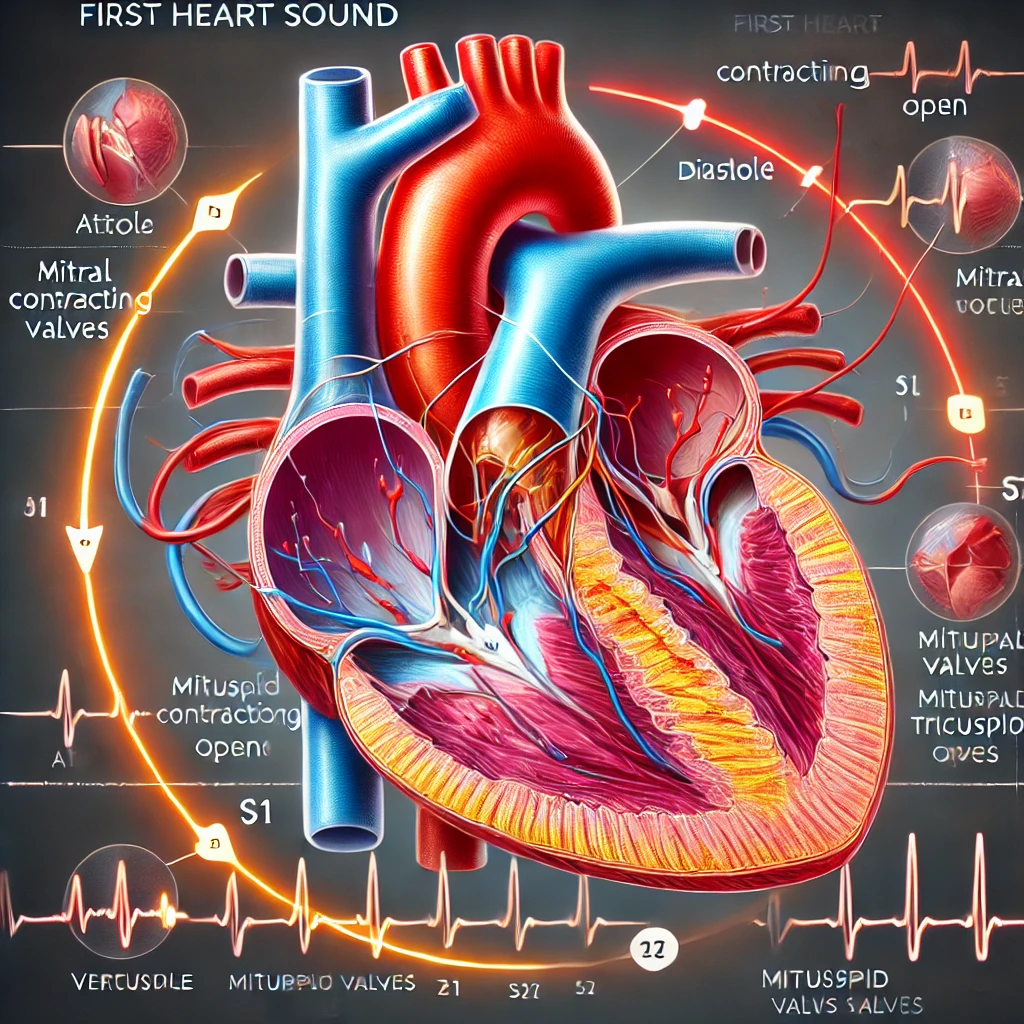
2.1.2 Heart Valves
Ensuring the one-way flow of blood through the heart are four heart valves, each located between the chambers and the large blood vessels connected to the core.
1. The Tricuspid Valve: Located between the right atrium and the right ventricle, it prevents blood from flowing back into the right atrium when the ventricle contracts.
2. The Pulmonary Valve: Located at the exit of the right ventricle, it prevents blood from flowing back into the ventricle after it is pumped out to the lungs.
3. The Mitral Valve: Located between the left atrium and the left ventricle, it prevents blood from flowing back into the left atrium when the ventricle contracts.
4. The Aortic Valve: Located at the exit of the left ventricle, it prevents blood from flowing back into the ventricle after it is pumped out to the body.
2.2 The Cardiovascular System
The heart is the central component of the cardiovascular system, which also includes the blood vessels—arteries, veins, and capillaries. The heart and blood vessels work together to circulate blood, delivering oxygen, nutrients, hormones, and other vital substances to the cells throughout the body. This continuous circulation also removes waste products like carbon dioxide and metabolic by-products.
2.2.1 The Role of Arteries and Veins
Arteries carry oxygen-rich blood from the heart to the body’s tissues, except the pulmonary artery, which carries oxygen-poor blood from the heart to the lungs for oxygenation. Veins carry oxygen-poor blood back to nature, except for the pulmonary veins, which carry oxygen-rich blood from the lungs to the heart.
2.3 Heart Wall Structure
The heart wall is composed of three layers:
1. Endocardium: The innermost layer, lining the heart chambers and valves. It’s composed of endothelial cells and connective tissue.
2. Myocardium: The thick middle layer comprises cardiac muscle cells. This layer contracts, creating the force to pump blood through the body.
3. Epicardium: The outermost layer, which also forms the inner layer of the pericardium, is a protective sac that encloses the heart.
2.4 The Electrical System of the Heart
The heart also contains a complex electrical system that controls the rate and rhythm of the heartbeat. This system includes the sinoatrial node (SA node), the atrioventricular node (AV node), and specialized conduction pathways (including the bundle of His and the Purkinje fibers) that coordinate the contraction of the heart chambers.
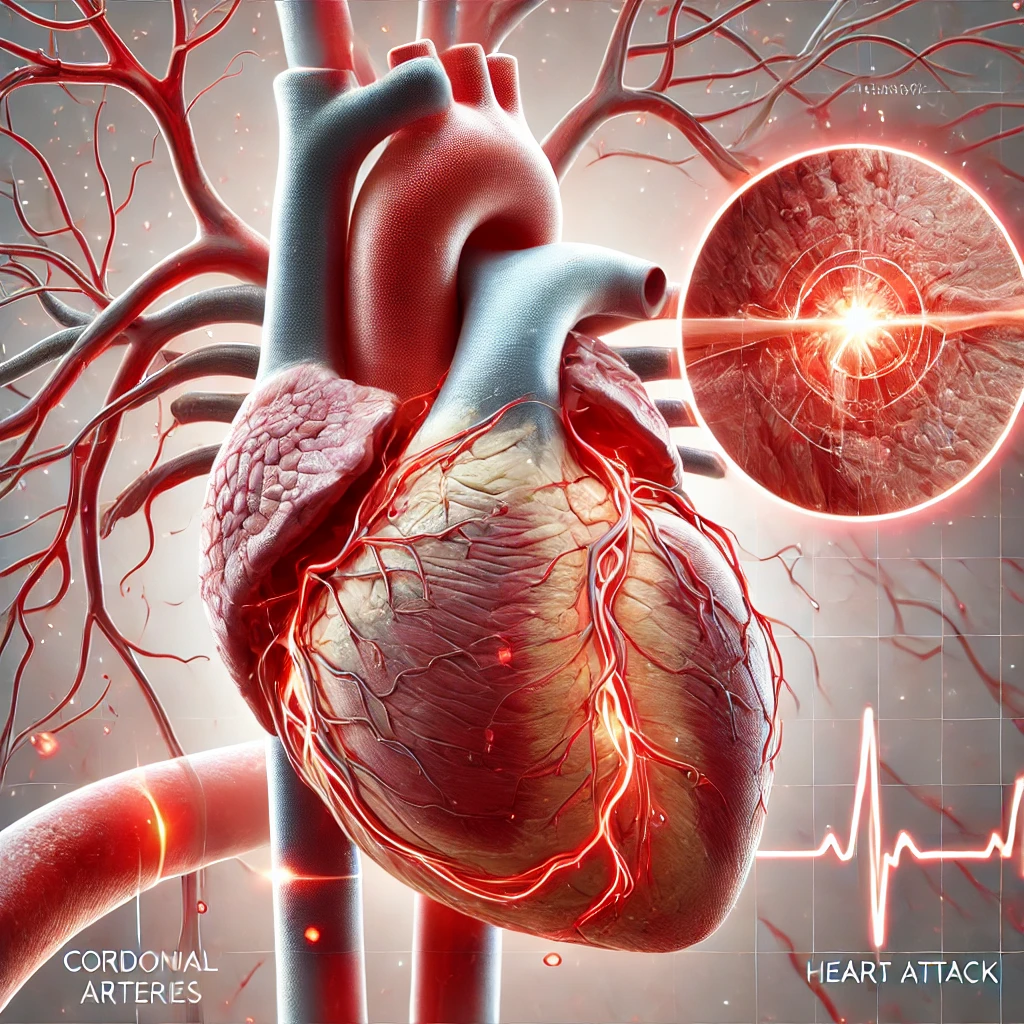
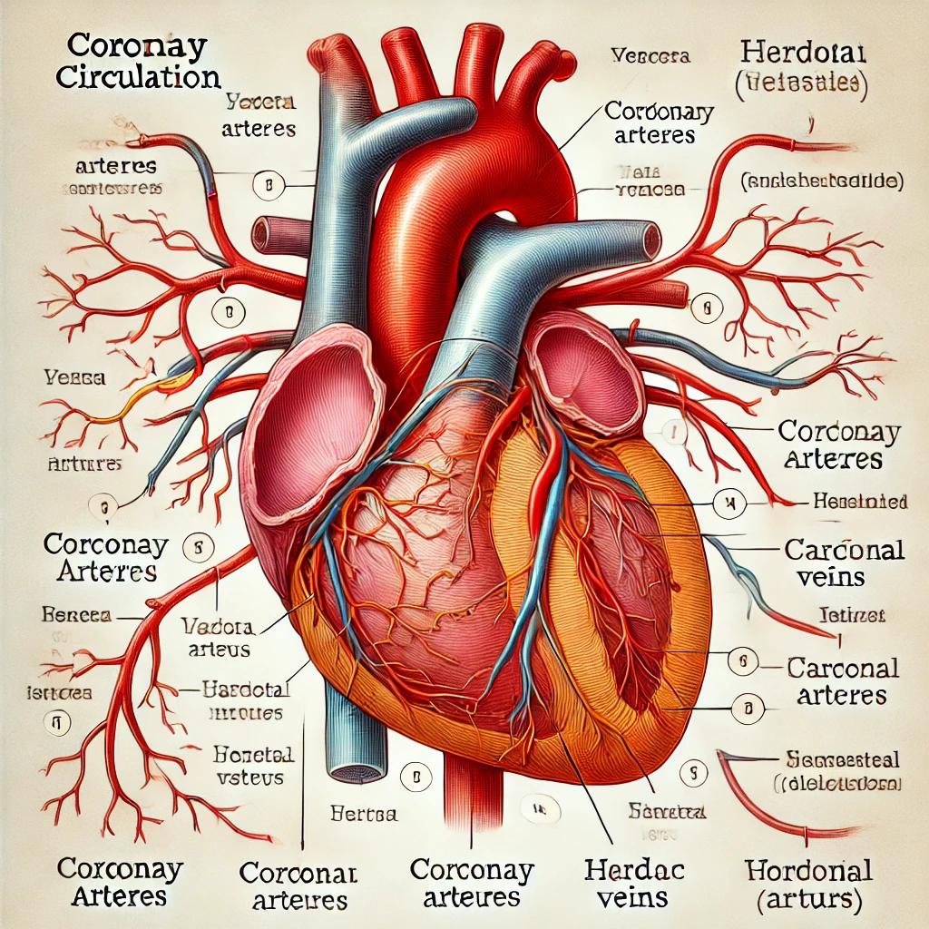
2.5 The Coronary Circulation
The heart muscle, like all tissues in the body, requires a supply of blood to provide it with oxygen and nutrients. This is provided by the coronary arteries, which branch off from the aorta just after it leaves the heart. After supplying the heart tissue, blood is returned to the right atrium through the coronary veins.
The complexity of the heart’s structure allows it to perform its vital function efficiently and without conscious effort. However, the effects can be profound and life-altering when something disrupts this precise structure – as in the case of congenital heart defects like the Tetralogy of Fallot. Understanding these structures and how they function paves the way for understanding how Tetralogy of Fallot disrupts the normal functioning of the heart.
4. Causes of Tetralogy of Fallot
Understanding the causes of Tetralogy of Fallot (TOF) requires a delve into the intricate world of human genetics and developmental biology. Despite being the most common form of cyanotic congenital heart disease, the exact reasons some babies are born with TOF remain largely unknown. However, current scientific understanding attributes the occurrence of TOF to a combination of genetic, environmental, and possibly random factors that affect heart development during the early stages of pregnancy.
4.1 Genetic Influences
In recent years, strides have been made in understanding the genetic factors contributing to TOF. While most cases appear sporadic (occurring by chance), studies suggest a familial predisposition in some instances, indicating genetic factors might play a role in Tetralogy of Fallot Causes.
TOF has been associated with several chromosomal abnormalities, gene mutations, and syndromes. For instance, an association has been found between TOF and the chromosomal disorder Down syndrome caused by an extra copy of chromosome 21. Other genetic conditions linked with TOF include DiGeorge syndrome (22q11.2 deletion syndrome), Alagille syndrome, and CHARGE syndrome.
Despite these links, no single gene has been pinpointed as a definitive cause of TOF in most cases, suggesting that the condition is likely the result of complex interactions among multiple genes and environmental factors.
4.2 Environmental Triggers
Certain environmental factors and maternal health conditions have been identified as potential risk factors for TOF. Pregnant women exposed to certain environmental toxins, such as pesticides or chemicals, may have an increased risk of having a child with TOF.
Additionally, maternal health conditions like poorly controlled diabetes and phenylketonuria can increase the risk. Pregnant women who suffer from poor nutrition, are obese, smoke, consume alcohol, or use illicit drugs are also at an increased risk. Certain medications taken during pregnancy, particularly anti-seizure medications, have also been implicated.
4.3 The Role of Chance
Given that most TOF cases occur sporadically with no apparent cause, some researchers believe that random events during the development of the heart in the fetus may lead to the condition.
In the developing fetus, the heart begins to form shortly after conception and continues to develop throughout pregnancy. TOF results from specific defects during the first eight weeks of fetal development, a critical period when the heart forms its chambers and related structures.
For reasons not entirely understood, the growth and division of cells in the heart can sometimes go awry during this stage, resulting in the abnormalities of TOF.
In conclusion, the causes of the Tetralogy of Fallot are multifaceted and still not entirely understood. Likely, a combination of genetic predisposition, environmental influences, and possibly chance events during heart development all contribute to this complex heart condition. Research continues to unravel the mystery behind the causes of TOF, hoping to find more effective ways to predict, prevent, and treat this condition.
4.4 Maternal Lifestyle Factors
Various maternal lifestyle factors during pregnancy have been associated with an increased risk of congenital heart defects, including the Tetralogy of Fallot. These factors encompass diet, substance use, and overall physical health.
Diet and Nutrition
Nutrition during pregnancy can significantly impact fetal development, and a diet low in certain essential nutrients can increase the risk of congenital heart defects. For instance, folate (or folic acid), a type of B vitamin, has been found crucial for preventing defects in the heart and other parts of the body during early embryonic development. Pregnant women who do not consume enough folic acid are at a higher risk of having a child with TOF.
Substance Use
The use of harmful substances like tobacco, alcohol, and illicit drugs during pregnancy is linked to an increased risk of various congenital disabilities, including TOF. Smoking during pregnancy, for example, affects oxygen and nutrient supply to the developing fetus, potentially resulting in heart defects. Alcohol use can also cause fetal alcohol syndrome, including heart defects like TOF.
4.5 Medical Conditions During Pregnancy
Certain medical conditions in the mother can increase the risk of having a child with TOF.
Maternal Diabetes
Both type 1 and type 2 diabetes, if poorly controlled, have been associated with a higher risk of congenital heart defects. High blood sugar levels can affect fetal heart development and lead to heart defects, including TOF.
Phenylketonuria (PKU)
PKU is a rare inherited disorder that increases phenylalanine in the blood. Mothers with PKU who don’t follow a low-phenylalanine diet during pregnancy have a higher chance of having a baby with TOF and other heart defects.
4.6 Medications During Pregnancy
Certain medications, when taken during pregnancy, have been associated with an increased risk of TOF. These include certain epilepsy medications, anti-inflammatory drugs, and specific acne treatments. Always consult with your doctor about the safety of drugs during pregnancy.
4.7 Advanced Maternal Age
Older maternal age, typically 35 and older, has been associated with a slightly increased risk of having a baby with a congenital heart defect, including TOF.
4.8 Syndromic Factors
While most cases of Tetralogy of Fallot occur sporadically, specific syndromes are known to increase the risk of TOF. These are usually genetic syndromes that affect multiple body systems.
DiGeorge Syndrome (22q11.2 Deletion Syndrome)
A deletion in a small part of chromosome 22 causes this genetic disorder. The DiGeorge syndrome affects many body systems and can cause various health problems, ranging from heart defects such as TOF to immune system issues, facial abnormalities, and developmental delays.
Down Syndrome (Trisomy 21)
Down syndrome is a genetic disorder caused by the presence of all or part of a third copy of chromosome 21. While Down syndrome is often associated with physical growth delays and intellectual disabilities, it can also increase the risk of congenital heart defects like TOF.
Alagille Syndrome
This genetic disorder can affect the liver, heart, and other parts of the body. One of the major features of Alagille syndrome is a liver disease caused by abnormalities in the bile ducts. However, heart problems, including TOF, can also occur in individuals with this condition.
CHARGE Syndrome
CHARGE syndrome is a complex genetic syndrome causing multiple congenital disabilities throughout the body. The name “CHARGE” is an acronym for some of the standard features of the condition: coloboma, heart defects, atresia choanae, growth retardation, genital abnormalities, and ear abnormalities. Heart defects associated with this syndrome may include TOF.
It’s important to note that while these syndromes are associated with an increased risk of TOF, most individuals with these conditions will not have TOF. Moreover, most cases of TOF are not related to these syndromes. These are merely risk factors, and having a risk factor does not guarantee the development of the condition.
4.9 Random Occurrence
In many cases, TOF occurs randomly, with no apparent cause. This suggests that in addition to the known genetic and environmental risk factors, other unknown factors may contribute to this condition’s development. Furthermore, it underscores the complexity of TOF and our incomplete understanding of its causes.
4.10 Closing Note on Causes of TOF
Although our understanding of the causes of Tetralogy of Fallot has improved, it remains incomplete. The condition is likely due to a complex interplay of various genetic, environmental, and possibly random factors. This understanding informs the prevention strategies and potential interventions to reduce the risk of TOF. However, in most cases, parents could not have done anything to prevent this condition. Understanding the causes helps healthcare professionals provide better care and guidance to families dealing with TOF. Continued research is essential to understand these factors further and improve our ability to prevent, diagnose, and treat this condition.
4.11 Maternal Exposure to Environmental Pollutants
Pregnant women exposed to certain environmental pollutants may be at an increased risk of having a baby with TOF. These pollutants can include industrial chemicals, pesticides, and certain metals.
Industrial Chemicals
Some industrial chemicals can interfere with the normal development of a fetus if a pregnant woman is exposed to them. For example, solvents used in dry cleaning and specific manufacturing processes have been linked to an increased risk of congenital heart defects.
Pesticides
Pesticides are widely used in agriculture and pest control. Some studies suggest that exposure to certain pesticides during pregnancy can increase the risk of having a baby with a congenital heart defect, such as TOF.
Metals
Exposure to certain metals, such as lead, during pregnancy can also increase the risk of congenital heart defects. Information can interfere with the normal development of an embryo or fetus and is toxic to many of the body’s systems.
4.12 Viral Infections During Pregnancy
Certain viral infections during pregnancy can increase the risk of congenital heart defects like TOF. For instance, if a woman contracts rubella (German measles) during pregnancy, especially during the first trimester, it can lead to several congenital disabilities, including TOF.
4.13 Maternal Stress and Mental Health
Emerging research suggests that high-stress levels and poor mental health in the mother during pregnancy may slightly increase the risk of having a baby with a congenital heart defect. However, this area of research is relatively new, and more studies are needed to confirm these findings and to understand the underlying mechanisms.
4.14 Additional Risk Factors
There may be additional risk factors for Tetralogy of Fallot that are not yet fully understood. Some studies have found associations between TOF and factors such as maternal obesity, maternal use of certain dietary supplements, and paternal smoking. However, more research is needed to confirm these associations and understand their significance.
4.15 Conclusion on Causes of Tetralogy of Fallot
The causes of Tetralogy of Fallot are complex and multifactorial. Likely, it results from a combination of genetic, environmental, and possibly random factors. Understanding these causes and risk factors can help develop prevention and early detection strategies. However, it’s crucial to note that having a risk factor does not guarantee that TOF will occur. Also, it’s possible to have TOF without these risk factors. Further research is required to understand this complex condition’s causes better and develop more effective treatments and interventions.