Tetralogy of Fallot, a congenital heart defect, presents a set of four related anomalies that significantly affect the structure of a newborn’s heart. Its diagnosis usually happens during infancy or sometimes before birth. This article sheds light on the various diagnostic procedures and tests undertaken to confirm the presence of Tetralogy of Fallot accurately.

Physical Examination
A physical examination is typically the first step in the diagnosis process. The healthcare provider will thoroughly examine the child, paying particular attention to the heart. A distinct heart murmur, which is an unusual sound produced due to turbulent blood flow in the heart, is often a clear indicator of a potential heart defect. This murmur may sound like a whooshing or swishing noise heard between heartbeats and is generally picked up while the doctor listens to the heart using a stethoscope.
Pulse Oximetry
Pulse oximetry is a simple, painless test used to measure the oxygen saturation level in the blood. It works by transmitting light through a thin part of the patient’s body, such as a finger or an earlobe. The light detector on the other side then measures the amount of light absorbed by the oxygen-carrying blood. Low oxygen saturation levels can indicate a heart defect, as the heart may not be effectively pumping oxygenated blood to the rest of the body.
Echocardiogram
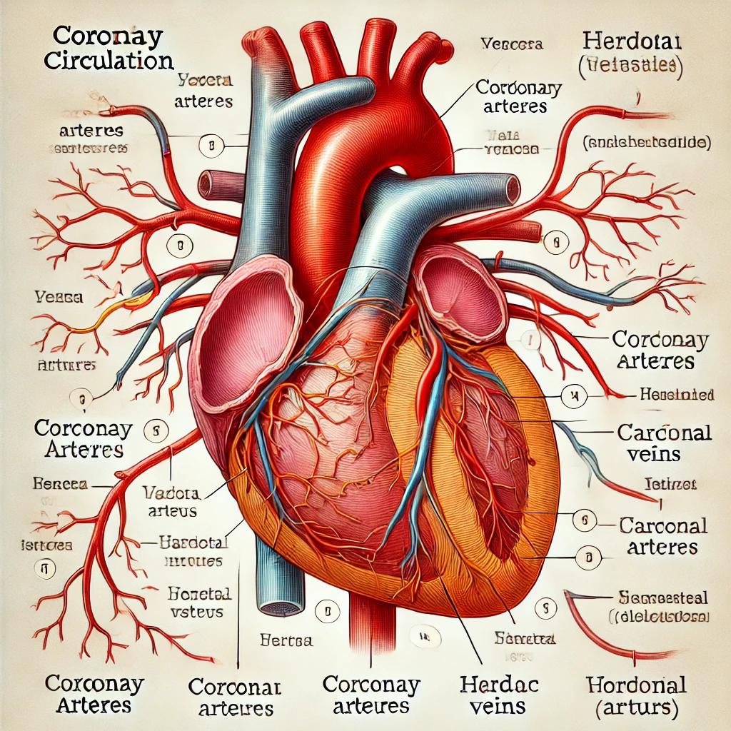
An echocardiogram is a non-invasive ultrasound of the heart. It uses high-frequency sound waves that bounce off the heart, creating detailed images of the heart’s structure. This allows doctors to clearly see any abnormalities or defects in the heart’s chambers and valves. In the case of Tetralogy of Fallot, doctors will look for the four specific heart defects that characterize this condition: a ventricular septal defect, pulmonary stenosis, right ventricular hypertrophy, and an overriding aorta.
Electrocardiogram (ECG)
An electrocardiogram, or ECG, measures the heart’s electrical activity. It involves attaching small sensor pads to the skin of the chest, arms, and legs. These sensors detect the electrical signals that control the rhythm of your heartbeat. Any abnormal patterns or rhythms can indicate heart problems. Specific patterns on an ECG can suggest Tetralogy of Fallot, such as a rightward shift in the heart’s electrical axis or signs of right ventricular hypertrophy (thickening of the heart muscle).
Chest X-Ray
A chest X-ray can provide a clear picture of the heart and lungs. This image can help doctors identify any unusual shapes or sizes in these areas. In the case of Tetralogy of Fallot, the heart may appear “boot-shaped” due to the right ventricular hypertrophy and the enlargement of the right atrium.
Cardiac MRI
Cardiac magnetic resonance imaging (MRI) is an advanced imaging technique that uses powerful magnets and radio waves to create detailed images of the heart. This test can provide a more detailed view of the heart’s structure and function than an echocardiogram, and it can confirm the diagnosis of Tetralogy of Fallot. It also helps doctors plan surgeries or other treatments by clearly showing the heart’s anatomy.
Cardiac Catheterization
Cardiac catheterization is an invasive procedure that can provide detailed information about the structures within the heart. During this procedure, a thin flexible tube (catheter) is inserted into a vein or artery, usually in the groin, and guided to the heart using X-ray imaging. The catheter can measure pressure and oxygen levels in different parts of the heart, helping to confirm the diagnosis of Tetralogy of Fallot and the severity of the condition.
Genetic Testing
As Tetralogy of Fallot can be associated with certain genetic conditions like Down syndrome and DiGeorge syndrome, genetic testing might be recommended to identify any such conditions. Genetic testing involves analyzing small samples of blood or body tissues to determine if you carry genes that could cause or increase the risk of certain medical conditions. It can help determine if the child’s condition is part of a larger genetic syndrome that could affect other parts of their health or their future family planning.
Remember, if your child shows any symptoms of Tetralogy of Fallot, such as cyanosis (bluish skin), difficulty feeding, or a heart murmur, you should seek medical attention immediately. Early diagnosis and intervention are key to managing this condition and improving your child’s long-term health outcomes.
References:
- Mayo Clinic – Tetralogy of Fallot
- Johns Hopkins Medicine – Tetralogy of Fallot
- American Heart Association – Tetralogy of Fallot
- Cleveland Clinic – Tetralogy of Fallot
- Stanford Children’s Health – Tetralogy of Fallot
You can read about the-
Disclaimer: The information contained on this site is intended for educational purposes only and is not a substitute for advice, diagnosis, or treatment by a licensed physician. It is not meant to cover all possible precautions, drug interactions, circumstances, or adverse effects. You should seek prompt medical care for any health issues and consult your doctor before using alternative medicine or making a change to your regimen.