Introduction to the Heart’s Electrical System
Before delving into the intricacies of the heart’s electrical conduction system, it is vital to grasp the fundamental role of the heart itself. The heart is a mighty organ and plays a crucial role in the circulatory system. This muscular organ, roughly the size of a fist, lies in the center of the chest, slightly tilted to the left. Its primary role is to act as a pump, propelling blood throughout the body to ensure the delivery of oxygen and nutrients to various tissues and organs. Additionally, the heart aids in the removal of metabolic waste products, including carbon dioxide, which is expelled from the body via the lungs.
The heart comprises four chambers: the right atrium, right ventricle, left atrium, and left ventricle. Each chamber plays a distinct role in the circulation of blood. The heart works in a dual system called the pulmonary and systemic circulations. In pulmonary circulation, the heart pumps deoxygenated blood from the body (via the right atrium and ventricle) to the lungs for oxygenation. The oxygenated blood then returns to the heart and enters the left atrium.
Next is systemic circulation, where the oxygen-rich blood from the left atrium is pumped into the left ventricle and then expelled from the heart to supply the entire body. After delivering oxygen and collecting waste products, the now-deoxygenated blood returns to the heart, and the cycle repeats.
This blood flow cycle within the body is continuous and ensures all body parts receive the necessary nutrients for proper function. It’s important to note that the heart’s ability to pump blood efficiently lies in its unique muscular structure and, most significantly, in its electrical conduction system, which triggers and coordinates the heart’s contractions. In the following sections, we will delve deeper into the workings of this fascinating electrical system, illuminating how it regulates the rhythmical beating of our hearts.
Overview of the Heart’s Electrical System
Following our exploration of the heart’s function, we now focus on its electrical conduction system, which plays a fundamental role in the synchronized contractions that make the heart an efficient and life-sustaining pump. As integral as it is intricate, this electrical system comprises specialized cardiac cells that generate and transmit electrical impulses throughout the heart, dictating its rhythmic beat.
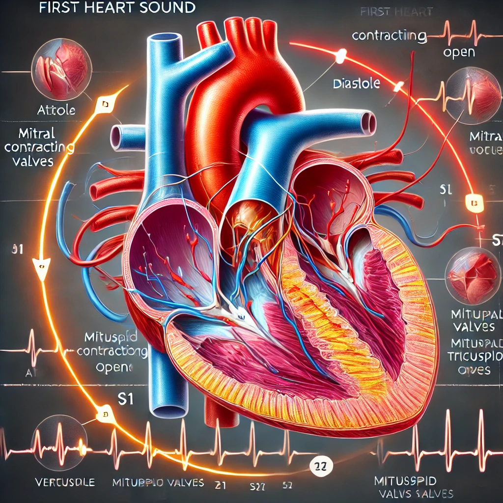
The heart’s electrical system is a natural pacemaker and conduction pathway. It ensures that each section of the heart contracts at the right time and sequence, creating the familiar “lub-dub” sound and effectively pumping blood to the lungs and the rest of the body.
The system initiates electrical impulses in the sinoatrial (SA) node, often dubbed the heart’s natural pacemaker due to its setting the heartbeat’s pace. Located in the right atrium, the SA node spontaneously generates electrical signals that radiate through the atria, causing them to contract and push blood into the ventricles.
The next stage in this electrical journey involves the atrioventricular (AV) node. The impulses, having induced atrial contraction, converge at the AV node, nestled at the junction of the atria and ventricles. Here, the signals undergo a brief delay, a critical pause that allows the ventricles to fill with blood before they contract.
Following this delay, the electrical impulses dart down the Bundle of His, a bundle of conductive fibers embedded in the heart’s septum. This bundle soon forks into the left and right bundle branches, which carry the electrical signals down to the apex of the heart.
Finally, these branches further divide into a network of finer fibers called Purkinje fibers. These fibers spread the impulses across the ventricular muscle, causing it to contract almost simultaneously and powerfully pump blood from the heart to the lungs and the rest of the body.
Importance of the Heart’s Electrical System
The importance of the heart’s electrical conduction system cannot be overstated. This intricate network serves as the heart’s own ‘internal pacemaker,’ responsible for coordinating and synchronizing the rhythmic contractions of the heart muscle that allow blood to circulate throughout the body.
Perhaps the most crucial aspect of the heart’s electrical system is its ability to maintain a regular, controlled heartbeat. An average human heart beats approximately 60 to 100 times per minute at rest, translating to more than 100,000 heartbeats daily. This constant rhythm ensures the continuous, uninterrupted delivery of oxygen-rich blood to all body parts. Without this regular rhythm, the organs and tissues would not receive the oxygen and nutrients they need to function correctly, leading to potential organ failure and death.
The electrical conduction system also ensures that the heart’s four chambers work together in a highly organized manner. For efficient blood flow, it is essential that the atria (the two upper chambers) contract first, followed by the ventricles (the two lower chambers). The delay introduced by the AV node allows the atria to fully empty their blood into the ventricles before the ventricles contract, maximizing the volume of blood pumped to the body with each heartbeat.
The heart’s electrical system is also responsible for responding to changes in the body’s demands for blood flow. For instance, during physical exercise or emotional Stress, the body requires more oxygen, increasing the heart rate. This change in heart rate is triggered by signals from the brain to the SA node, demonstrating how the heart’s electrical system interacts with the nervous system to adapt to changing conditions.
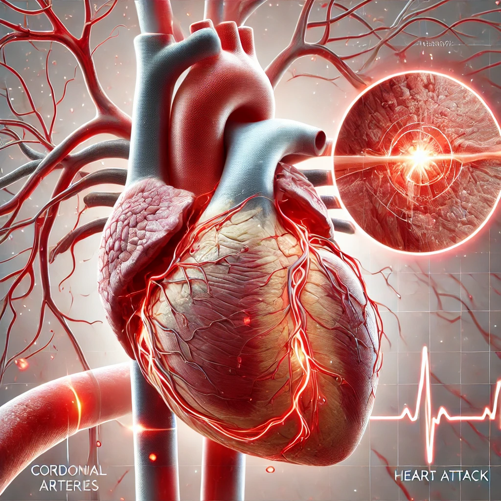
It’s also noteworthy that disturbances or malfunctions in the heart’s electrical system can lead to arrhythmias characterized by irregular heartbeat. Arrhythmias can affect the heart’s ability to pump blood effectively, leading to various symptoms and potentially severe health complications. Therefore, understanding the heart’s electrical system is crucial for comprehending the heart’s normal function and diagnosing and treating a range of heart conditions.
Overall, the heart’s electrical system is essential for maintaining life. It ensures the heart beats in a consistent, coordinated manner, enabling the efficient circulation of blood to all parts of the body. It also facilitates the heart’s response to changing conditions, demonstrating its vital role in health and disease. In the following sections, we will examine the individual components of this electrical system in greater detail.
The Components of the Heart’s Electrical System

Sinoatrial (SA) Node
The Sinoatrial (SA) Node is a small, crescent-shaped mass of specialized cardiac cells located in the upper part of the right atrium, near the opening of the superior vena cava. Often referred to as the heart’s “natural pacemaker,” the SA Node is crucial in initiating each heartbeat.
Unlike most other cells in the body, cells in the SA Node can depolarize and spontaneously generate an electrical impulse. This automaticity allows the SA Node to set the heart’s rhythm. Under normal conditions, the SA Node generates an electrical impulse approximately 60 to 100 times per minute, establishing what’s known as the normal sinus rhythm.
The electrical impulse generated by the SA Node spreads rapidly through the walls of the right and left atria, causing them to contract almost simultaneously. This contraction pushes the blood from the atria into the lower chambers of the heart, the ventricles, which are relaxed at this point.
The electrical activity of the SA Node can be influenced by the autonomic nervous system, which comprises the sympathetic and parasympathetic nervous systems. The sympathetic nervous system, often associated with the ‘fight or flight’ response, increases heart rate, while the parasympathetic nervous system, associated with the ‘rest and digest response, slows it down. This balance lets the heart respond dynamically to the body’s changing oxygen and nutrient-rich blood needs.
Dysfunction of the SA Node can lead to various arrhythmias, including sinus bradycardia (a prolonged heart rate), sinus tachycardia (an abnormally fast heart rate), or sinus arrhythmia (irregular heartbeat), among others. While some conditions can be asymptomatic or benign, others may require medical intervention.
In the subsequent sections, we will explore how the electrical impulse generated by the SA Node propagates to the other parts of the heart, starting with the Atrioventricular (AV) Node, the next major component of the heart’s electrical conduction system.
Atrioventricular (AV) Node
The Atrioventricular (AV) Node is another critical component of the heart’s electrical conduction system. Situated at the lower part of the right atrium, close to the ventricles, it plays a vital role in maintaining the orderly sequence of heart contractions.
After the Sinoatrial (SA) Node fires an electrical impulse that causes the atria to contract, this impulse arrives at the AV Node. Here, the electrical signal is briefly delayed before it is allowed to pass into the ventricles. This delay, usually around 0.1 seconds, is crucial as it allows the atria enough time to fully empty their blood content into the ventricles before the ventricles contract. This ensures that the maximum volume of blood is delivered into the ventricles to be pumped out to the body and lungs, thereby maximizing the efficiency of each heartbeat.
The AV Node is the only electrical connection between the atria and the ventricles. Suppose it fails or gets blocked (a condition known as heart block). In that case, the atria and ventricles can beat independently, leading to less efficient blood pumping and potentially causing symptoms such as dizziness, shortness of breath, or fainting.
After passing through the AV Node, the electrical impulses travel along the Bundle of His, which is the following structure in the heart’s electrical conduction system. Like the SA Node, the AV Node’s activity can also be influenced by the autonomic nervous system, with sympathetic stimulation increasing its rate of conduction and parasympathetic stimulation slowing it down.
Overall, the AV Node plays an essential role in the heart’s electrical system, ensuring the proper timing of contractions between the atria and the ventricles. Its dysfunction can lead to various types of heart block, potentially requiring medical intervention such as a pacemaker, a device that can artificially regulate the heart’s rhythm. The following sections discuss the Bundle of His and its role in the heart’s electrical conduction system.
Bundle of His (AV Bundle)
The Bundle of His, also known as the Atrioventricular (AV) Bundle, is an essential part of the heart’s electrical conduction system. It is named after Wilhelm His Jr., the Swiss cardiologist who first described it.
The Bundle of His is located at the top of the interventricular septum, the wall that separates the heart’s two ventricles. It acts as a highway, transmitting the electrical impulses from the Atrioventricular (AV) Node into the ventricles.
After the electrical impulse passes through the AV Node and experiences the brief delay necessary to ensure the atria have finished contracting, it enters the Bundle of His. The Bundle of His doesn’t delay the impulse any further; instead, it provides a rapid, direct path for the impulse to reach the ventricles.
An essential characteristic of the Bundle of His is that it is the only electrical connection between the atria and the ventricles. This one-way traffic ensures that the electrical impulses move only in the intended direction, from the atria to the ventricles, and not in reverse.
At the end of the Bundle of His, the pathway splits into two branches, known as the right and left bundle branches. These branches carry the electrical impulse to the right and left ventricles. The electrical impulse causes the ventricles to contract, pushing blood out of the heart and into the lungs and the rest of the body.
A blockage or delay in the Bundle of His can cause what’s known as a bundle branch block. This can slow down or interrupt the flow of electrical impulses to the ventricles, causing an irregular or abnormal heartbeat. Depending on the location and severity of the block, treatment may be required to restore normal heart rhythm.
In summary, the Bundle of His is a critical component of the heart’s electrical system, ensuring that the electrical impulses move efficiently from the atria to the ventricles. In the subsequent sections, we will examine the right and left bundle branches and their role in this unique system.
Right and Left Bundle Branches
The right and left bundle branches follow the Bundle of His in the heart’s electrical conduction system. These structures are extensions of the Bundle of His and act as electrical pathways leading into the heart’s lower chambers, the ventricles.
As the electrical impulse travels down the Bundle of His, it splits into the right and left bundle branches. These branches run down the sides of the interventricular septum, the muscular wall that separates the two ventricles. They serve to rapidly transmit the electrical impulse from the Bundle of His to the ventricular muscle.
The right bundle branch carries the electrical impulse to the right ventricle, causing it to contract and pump blood to the lungs via the pulmonary artery. This is where the blood gets oxygenated before returning to the heart’s left atrium.
Simultaneously, the left bundle branch, which further divides into the left anterior and posterior fascicles, delivers the electrical impulse to the left ventricle. The left ventricle pumps oxygen-rich blood to the rest of the body via the aorta, the largest artery.
The right and left bundle branches play a critical role in synchronizing the contraction of the right and left ventricles. This ensures that blood is efficiently pumped from the heart to the lungs and the rest of the body.
A blockage in one or both bundle branches can lead to a condition known as a bundle branch block. This can cause the corresponding ventricle to contract later than usual, leading to a weak heartbeat. Depending on the extent and location of the block, this condition may cause no symptoms or lead to a noticeable irregular heartbeat or even more severe cardiac issues.
The right and left bundle branches ensure that the electrical impulse reaches all parts of the ventricles, facilitating their coordinated contraction and the effective pumping of blood. The Purkinje fibers follow these bundle branches in the heart’s electrical conduction system, which we will discuss in the next section.
Purkinje Fibers
The Purkinje fibers represent the final step in the heart’s electrical conduction system, transmitting the electrical impulses that trigger the ventricles’ contraction to the farthest reaches of the heart muscle. Named after the Czech anatomist Jan Evangelista Purkinje discovered them, these fibers are unique and specialized cardiac cells.
Located within the walls of the ventricles, the Purkinje fibers branch out from the right and left bundle branches, extending like a complex network of fine threads throughout the ventricular muscle. Their primary function is to rapidly conduct the electrical impulses from the bundle branches to the ventricular myocardium, the muscular layer of the heart wall.
This rapid conduction of the electrical impulse ensures that the contraction of the ventricles is almost simultaneous, promoting a more efficient and robust heartbeat. It allows the right ventricle to pump blood into the lungs to pick up oxygen while the left pumps oxygenated blood to the rest of the body.
Notably, the Purkinje fibers can also generate electrical impulses on their own, albeit slower than the SA node. This characteristic serves as a backup mechanism: if the SA node or other parts of the conduction pathway fail to generate impulses, the Purkinje fibers can take over to keep the heart beating, although this would result in a slower heart rate.
Abnormalities in the Purkinje fibers can lead to certain types of arrhythmias. These include premature ventricular contractions, or PVCs, which are extra, abnormal heartbeats originating in the ventricles, disrupting the regular heart rhythm.
In summary, the Purkinje fibers are integral to the heart’s electrical conduction system, ensuring that the electrical impulses efficiently reach all parts of the ventricles. The entire conduction system, from the SA node to the Purkinje fibers, works harmoniously to orchestrate the rhythmic contractions of the heart, thereby enabling the continuous and efficient circulation of blood throughout the body.
The Process of Electrical Conduction in the Heart

The Generation of Electrical Impulses in the SA Node
The generation of electrical impulses in the Sinoatrial (SA) Node is an intricate process that lies at the heart of the body’s circulatory function. These impulses set the pace for the heart’s rhythmic contractions, thereby ensuring the efficient circulation of blood throughout the body.
Cells in the SA Node, unlike most cells in the body, possess automaticity – the unique ability to generate electrical impulses without any external trigger spontaneously. This property enables the SA Node to function as the heart’s natural pacemaker.
The process of generating an electrical impulse begins with the slow influx of sodium ions into the SA Node cells during the resting phase of the cardiac cycle. This slow, gradual influx of sodium ions is known as the ‘funny current’ or ‘pacemaker current.’ It increases the cell’s positive charge, bringing it closer to the threshold potential – the point at which an action potential, or electrical impulse, is generated.
When the threshold potential is reached, calcium ions are rapidly absorbed into the cell, while potassium ions start to leave the cell. This shift of ions changes the electrical charge within the cell, resulting in depolarization – the process that creates an electrical impulse.
Following depolarization, there is a rapid efflux of potassium ions out of the cell, which results in repolarization or the restoration of the cell’s negative charge. After repolarization, the cell enters a brief phase of hyperpolarization, where it is slightly more negative than at its resting potential. The slow influx of sodium ions starts again, and the cell slowly depolarizes, setting the stage for generating the next electrical impulse.
The entire process, from the slow influx of sodium ions to the rapid efflux of potassium ions, happens approximately 60 to 100 times per minute in a resting heart, setting the pace for the heart’s rhythm. Each electrical impulse the SA Node generates spreads rapidly across the atria, triggering their contraction and initiating a new heartbeat.
It’s important to note that the autonomic nervous system can influence the rate of impulse generation in the SA Node. The sympathetic nervous system can increase the heart rate, while the parasympathetic nervous system can decrease it, allowing the heart to adapt to the body’s changing needs.
In summary, generating electrical impulses in the SA Node is a complex, meticulously regulated process that ensures the heart beats consistently and coordinatedly, making it indispensable to the heart’s function and, by extension, to life itself.
Propagation of the Impulse to the AV Node and the Brief Delay
Once the electrical impulse is generated in the Sinoatrial (SA) Node, it must travel throughout the heart to stimulate the coordinated contractions that pump blood to the body and lungs. The first step of this journey is the impulse propagation to the Atrioventricular (AV) Node.
The SA Node is located in the upper part of the right atrium, and the AV Node is positioned in the lower part of the atrium, near the ventricles. The electrical impulse generated by the SA Node spreads rapidly across the walls of the atria, causing them to contract and push blood into the ventricles.
The atrial muscle cells are connected by intercalated discs containing gap junctions. These gap junctions allow the rapid and direct passage of ions between cells, effectively creating a continuous pathway for the electrical impulse to travel across the atria.
Upon reaching the AV Node, the electrical impulse experiences a crucial delay, often around 0.1 seconds. This delay is not due to the distance the impulse has traveled but is a unique feature of the AV Node itself, which has a slower rate of electrical conduction. This delay is critical for the heart’s functioning. It allows the atria to fully contract and empty their blood content into the ventricles before they start to contract.
Without this delay, the ventricles might begin contracting before they are filled with blood, reducing the volume of blood pumped out to the body and lungs, leading to less efficient circulation. Therefore, this brief delay at the AV Node is an essential part of the cardiac cycle, ensuring the heart operates in a coordinated and efficient manner.
In the next stage of the cardiac cycle, the electrical impulse travels from the AV Node down the Bundle of His and its branches before reaching the Purkinje fibers and stimulating the ventricles to contract. But it’s in this delay at the AV Node that we find the heartbeat’s rhythm, a moment of stillness before the powerful contraction of the ventricles sends blood coursing through our veins.
XI. Transmission of the Impulse Down the Bundle of His
After the crucial delay in the Atrioventricular (AV) Node, the electrical impulse continues its journey down the heart’s electrical conduction system via the Bundle of His. This structure plays a crucial role in transmitting the electrical impulses from the atria to the ventricles, ensuring the coordinated contraction of the heart muscle.
The Bundle of His, also known as the AV Bundle, is a thin band of fibers that originates from the AV Node and extends into the interventricular septum, the wall separating the right and left ventricles. The structure serves as the only electrical bridge between the atria and ventricles, signifying the importance of its role in the heart’s electrical system.
After traversing the delay at the AV Node, the electrical impulse enters the Bundle of His. Despite the previous delay, the conduction of impulses through the Bundle of His is rapid, ensuring a swift transition of the impulse to the ventricles. This rapid transmission is due to the unique properties of the cells within the Bundle of His, designed for efficient and high-speed conduction of electrical signals.
At the end of the Bundle of His, the pathway splits into two main branches, the right, and left bundle branches. These branches carry the electrical impulse to the right and left ventricles. The division ensures that the impulse spreads uniformly across both ventricles, leading to their simultaneous contraction.
However, transmitting the electrical impulse down the Bundle of His is not without potential issues. If blockage or impairment occurs in the Bundle of His, a condition known as heart block can ensue. This can lead to an irregular or slow heartbeat, as the electrical signals from the atria have difficulty reaching the ventricles. In some cases, treatment may be required to restore the normal conduction of impulses and maintain an effective heartbeat.
In essence, the transmission of the impulse down the Bundle of His is a critical stage in the heart’s electrical activity. This structure ensures the efficient and synchronized transfer of electrical impulses from the atria to the ventricles, enabling the heart to fulfill its function as the body’s primary blood pump.
XII. The Splitting of the Impulse Through the Right and Left Bundle Branches
The right and left bundle branches are extensions of the Bundle of His and form an integral part of the heart’s electrical conduction system. Their primary role is to carry the electrical impulse from the Bundle of His to the walls of the ventricles, triggering their contraction.
After the electrical impulse passes through the AV Node and the Bundle of His, it splits into two distinct pathways: the right and left. This bifurcation ensures that the impulse can reach both ventricles simultaneously.
The right bundle branch carries the electrical impulse along the right side of the interventricular septum, the muscular wall that separates the two ventricles, before spreading throughout the muscle tissue of the right ventricle. Once the impulse reaches the muscle cells of the right ventricle, it triggers their contraction, causing the right ventricle to pump deoxygenated blood to the lungs via the pulmonary artery.
Simultaneously, the left bundle branch transmits the electrical impulse to the left ventricle. The left bundle branch further divides into two installments – the left anterior fascicle and the left posterior fascicle – to distribute the impulse throughout the sizeable left ventricle. The left ventricle then contracts, pumping oxygenated blood to the rest of the body via the aorta, the body’s leading and largest artery.
The synchronized contractions of the ventricles, triggered by the simultaneous transmission of the electrical impulse through the right and left bundle branches, are crucial for effectively pumping blood. If the impulse doesn’t reach one ventricle at the same time as the other, it could result in a weak, irregular heartbeat. This makes the right and left bundle branches a critical component of the heart’s electrical conduction system.
Blockage or damage to one or both bundle branches can lead to a condition known as bundle branch block. This can slow or interrupt the flow of electrical impulses to the ventricles, potentially causing an irregular heartbeat or other cardiac issues.
In conclusion, splitting the impulse through the right and left bundle branches is a crucial step in the heart’s electrical activity, ensuring the coordinated and efficient contraction of the heart’s powerful lower chambers, the ventricles.
The Spreading of the Impulse by the Purkinje Fibers
The final and crucial step in the heart’s electrical conduction system involves the Purkinje fibers, a network of fine, conductive fibers that disperse the electrical impulse throughout the ventricular myocardium, triggering the coordinated contraction of the ventricles.
Originating from the left and right bundle branches, the Purkinje fibers fan out in a complex network across the inner ventricular walls. This extensive network ensures the electrical impulse reaches all parts of the ventricles. The speed and efficiency of this system are remarkable. Despite their far-reaching network, the Purkinje fibers distribute the electrical impulse nearly simultaneously to all parts of the ventricles. This results in a near-simultaneous contraction of the entire ventricular myocardium, which is crucial for effectively pumping blood.
The Purkinje fibers have a unique feature among the cells in the heart’s conduction system: they can initiate an electrical impulse independently, much like the SA Node, albeit at a much slower rate. This is a critical backup system. If the SA Node or any other part of the conduction pathway fails, the Purkinje fibers can generate their impulses to keep the heart beating, albeit slower and less efficiently.
Abnormalities or damage to the Purkinje fibers can lead to various cardiac arrhythmias. Since these fibers are responsible for the final propagation of the electrical impulse, any disruption in their function can result in uncoordinated or irregular ventricular contractions, leading to inefficient blood pumping.
In conclusion, the spreading of the electrical impulse by the Purkinje fibers represents the final step in the heart’s orchestrated symphony of electrical activity. Their extensive network ensures the fast and uniform contraction of the ventricles, enabling the effective ejection of blood into pulmonary and systemic circulation. This marks the end of one cardiac cycle and the beginning of the next, driven by the continuous, rhythmic dance of electrical activity in the heart.
Next:
Regulation of the Heart’s Electrical Activity
- Role of the autonomic nervous system
- Influence of hormones
- Effect of physical activity and Stress
References:
- American Heart Association – How the Healthy Heart Works
- National Institutes of Health – The Heart’s Electrical System
- British Heart Foundation – Your Heart’s Electrical System
- Harvard Medical School – Understanding the Heart’s Electrical System
- Cleveland Clinic – The Conduction System
My other Articles:
Disclaimer: This article provides general information about the heart’s electrical system. It does not constitute medical advice and should not be used to diagnose or treat health problems. Always consult with a qualified healthcare professional for medical advice. While every effort has been made to ensure the accuracy of this information, the author and publisher accept no responsibility for errors or omissions.